Innovative Approaches for 3D Image Analysis of Blood Vessels
2 March 2012
K. Rohr, Project QuantVessel
A research project on the development of innovative methods for three-dimensional (3D) image analysis of blood vessels will represent Heidelberg University and the German Cancer Research Center (DKFZ) at an exhibition of the German Research Foundation (DFG). Under the title “From an Idea to Discovery” („Von der Idee zur Erkenntnis“), the DFG presents outstanding scientific projects that are funded by the foundation in the German Bundestag (Federal Parliament) from 6 to 30 March 2012. The Heidelberg project “QuantVessel” is headed by associate professor Dr. Karl Rohr and carried out at the BioQuant Centre, the Institute of Pharmacy and Molecular Biotechnology of Heidelberg University and the Division Theoretical Bioinformatics of the DKFZ.
As Dr. Rohr explains, a challenge in the accurate measurement – quantification – of blood vessels is to capture the complex and curved 3D anatomy from 2D image slices. „Existing image analysis systems for automated quantification generally do not fulfil clinical requirements regarding their accuracy, and reliable solutions are lacking“, the scientist emphasizes. Dr. Rohr and his research group „Biomedical Computer Vision“ work particularly on the 3D quantification of the aorta to treat pathologic dilations, i.e. aneurysms.
Photo: Private
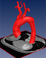
The scientists have developed a new 3D technique which accurately determines the size and shape of blood vessels from 3D tomographic image data, for example from computed tomography angiography (CTA). To this end, they combine so-called 3D intensity models with mathematically well-founded optimisation methods. The models describe the variation of images in the intensity values of blood vessels and are fitted to the image data of a particular person during the computer-based analysis in order to determine size and shape parameters, in particular, the diameter along vessels. This approach enables quantification of blood vessels with arbitrary orientation, different curvature and with branches.
As Dr. Rohr explains, the approach developed in Heidelberg is more accurate than previous approaches to image analysis, particularly with respect to determining the diameter of blood vessels. At the same time, additional geometric parameters like, for example, the length and curvature of the outer contour of the aortic arch can be determined. Moreover, the approach was extended to automatic analysis of time-resolved dynamic 4D images during the heart cycle. “The quantification results from image analysis are important to select optimal individual vessel prostheses (stent grafts) for operation planning“, Dr. Rohr says. The research work is carried out in close cooperation with radiologists and vascular surgeons of Heidelberg University Hospital and the German Cancer Research Center, in particular with Dr. Hendrik von Tengg-Kobligk as well as with Prof. Dr. Hans-Ulrich Kauczor and Prof. Dr. Dittmar Böckler. A clinical study is planned.
The exhibition “From an Idea to Discovery” will be opened on 6 March by the President of the Bundestag, Prof. Dr. Norbert Lammert, and the President of the German Research Foundation, Prof. Dr. Matthias Kleiner, and will be shown at the Paul-Löbe-Haus in Berlin until the end of March. The project “Quantification of the Morphology of Human Vessels from 3D Tomographic Image Data” (QuantVessel) will be presented in pictures, additional texts and videos, a 3D print of an aorta as well as the software programme developed for this approach especially. The project is one of ten projects on show at the DFG exhibition, selected from about 20,000 projects the DFG funds annually and the only one from the state of Baden-Württemberg.
The research group „Biomedical Computer Vision“ headed by Dr. Rohr develops computer science methods for automatic analysis of medical and biological images. Particularly participating in the project “QuantVessel“ are Dr. Stefan Wörz and Andreas Biesdorf as well as Simon Eck and Wei Liao. For more information go to www.bioquant.uni-heidelberg.de/research/groups/biomedical_computer_vision/research/quantvessel.html.
Information on the exhibition:
http://www.bundestag.de/kulturundgeschichte/ausstellungen/parl_hist/idee_erkenntnis/index.jsp
Note to news desks:
Digital pictures are available from the press office.
Contact:
PD Dr. Karl Rohr
Heidelberg University, BioQuant Centre and
Institute of Pharmacy and Molecular Biotechnology
German Cancer Research Center
Division Theoretical Bioinformatics
Phone: +49 6221 54 51298
k.rohr@uni-hd.de
Communications and Marketing
Press Office
Phone: +49 6221 542311
presse@rektorat.uni-heidelberg.de