

TP 5: :Role of APP and APPsα in denervation-induced plasticity in the hippocampus
 |
|
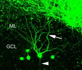
Figure Legend:
Organotypic slice culture of a Thy-1-GFP mouse (Feng et al., 2000). In the dentate gyrus, single granule cells are strongly GFP-labeled and individual granule cell dendrites (arrow) and axons (arrowhead) can be identified. Pharmacological and denervation experiments can be performed and the structural and functional changes of these cells can be studied over time using high-resolution time-lapse imaging. GCL - granule cell layer; ML - molecular layer. |
Following traumatic injury of the adult brain APP is upregulated at injury sites and in denervated brain regions (Murakami et al., 1998; Ramirez et al., 2001). Since APP and its fragment APPsα are involved in the regulation of synaptic plasticity (Anliker and Müller, 2006; Ring et al., 2007) and, furthermore, have neuroprotective effects in vitro (Mattson et al., 1993; Kögel et al., 2005; Copanaki et al., 2007; 2010; Eckert et al., 2011), we hypothesize that the induction of APP after brain injury could influence reorganization responses of surviving neurons. At present, however, the precise role of APP and its fragments under conditions of brain injury are unclear and whether or not they are relevant for brain repair needs to be determined.
In previous studies, the structural reorganization of denervated neurons in the brain was analyzed using the classical entorhinal denervation model (see Deller and Frotscher, 1997, for review). In recent years we have transferred this model to the in vitro situation (Del Turco and Deller, 2007; Müller et al., 2010), which makes it possible to visualize the reorganization of the dentate gyrus using time-lapse imaging techniques (confocal imaging; 2-photon-microscopy) and to perform functional studies using pharmacology and mouse mutant resources (e.g., Müller et al,. 1994). By using organotypic slice cultures of Thy1-GFP mice, which express GFP in only a subpopulation of neurons (Feng et al., 2000; Müller et al., 2010), the reorganization of single neurons can be studied. Specifically, we will address the following questions: (1) What is the effect of APP and APPsα on denervated dendrites? (2) What is the effect of APP and APPsα on sprouting axons? (3) Which signalling pathways are involved? (4) Can APPsα enhance lesion-induced reorganization? By addressing these questions we hope to gain novel insights into the physiological role of APP and APPsα in the normal and the injured brain.