

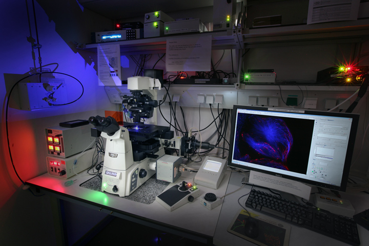
Nikon TIRF/FRAP microscope
The Nikon TIRF system with Nikon's H-TIRF module is an inverted and fully automated Ti2 microscope with objective TIRF illumination. A Nikon Perfect Focus System (PFS of the 4th generation) is included that continuously determines the distance to the coverslip and readjusts it if necessary (to counteract thermal drift or vibrations). For live cell imaging of mammalian cells, the system is equipped with an on-stage incubation chamber from TokaiHit, controlling temperature, CO2-concentration and humidity.
TIRF is short for Total Internal Reflection Fluorescence (Microscopy): By directing a laser beam at an very shallow angel towards the coverslip on which cells adhere (and on the condition that the refractive index of the medium on that side is lower than that of glass and oil), the light will not penetrate the coverslip but be totally reflected. Under this condition an ultra thin (around 100 to 200 nm) "evanescent electromagnetic field" of light forms on the side the cells are adhering. This means that in a TIRF microscope all fluorescently labelled structures of adhering cells as close as 100 to 200nm from the coverslip will become visible but everything further afar will stay black. In this region the cell membrane and the adjacent cytoskeleton are located and can be studied without being affected by extrafocal blur.
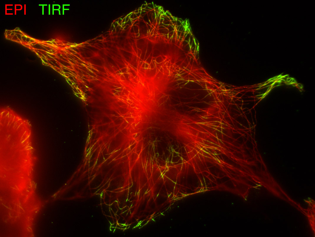
Comparison between TIRF (green) and widefield (red) image.
Source: NIC@Uni-HD
A confocal microscope would of course be able to show any image plane, not merely the one closest to the coverslip. But firstly a confocal could not selectively illuminate such a narrow slice (but rater something in the order of 300 to 400nm) and secondly a TIRF microscope does not need a confocal pinhole for imaging but is able to visualize the region of the cell membrane in total with a high sensitive EMCCD camera. Such cameras offer much higher quantum efficiencies compared to photomultipliers that are used as detectors in confocals. In the end, a TIRF microscope is limited to showing the region of the cell membrane but it can do so in much better quality and with much higher frame rates than a confocal microscope would be able to do.
Nikon's new H-TIRF Module, controlled by the imaging software NIS-Elements, allows auto alignment of settings, such as laser incident angles and evanescent wave field penetration depths. These parameters can be fine-tuned to best suit the needs of the individual user. Individual settings can be saved and recalled in order to easily repeat observations.In addition there is an Andor "FRAPPA" unit installed, for FRAP and photoactivation experiments like measurement of molecular recruitment rates, trafficking and turnover in single cells and sub-cellular organelles.
Objectives:
- Nikon Plan Fluor 10x NA 0.3 (working distance 16mm), corrected for 0.17mm coverslips (= type #1.5).
- Nikon Plan Fluor 20x NA 0.75 multi immersion (oil, glycerol, water; working distance 0.35 mm), corrected for 0.17mm coverslips (= type #1.5).
- Nikon S Fluor 40x NA 1.30 oil immersion objective (working distance 0.22 mm), corrected for 0.17mm coverslips (= type #1.5).
- Nikon Apo TIRF 60x NA 1.49 oil immersion objective (working distance 0.12mm) adjustable for glas coverslips with a thickness between 0.13 and 0.21mm), temperature adjustment for 23° and 37°C.
- Nikon Plan Apo λ 100x NA 1.45 oil immersion objective (working distance 0.13mm), corrected for 0.17mm coverslips (= type #1.5).
Please note: not all the objectives are included in the system permanently. Some are only given to the users on request.
Cameras
- Andor iXon Ultra DU-897U single photon detection EMCCD camera (for TIRF). This camera offers a resolution of 512 x 512 pixels with a gigantic on-chip pixel size of 16 x 16µm and a pixel well capacity of 180,000 electrons. The chip is cooled down to -100°C and 56 full frames can be read out per second.
- Andor Neo 5.5 sCMOS. The resolution of this camera is 2560 x 2160 pixels at a physical pixel size of 6.5 x 6.5μm.
Lasers for TIRF
- 405nm
- 488nm
- 561nm
- 640nm