
Motor Neurons Tell Blood Vessels Where To Go
30 March 2017
Source: Patricia Himmels and Carmen Ruiz de Almodóvar
Heidelberg Neuroscientists have identified a critical regulator for blood vessel growth in the developing embryonic spinal cord. The research group under the direction of Dr Carmen Ruiz de Almodóvar of the Heidelberg University Biochemistry Center discovered that special nerve cells known as motor neurons control this process. This new insight into the nature of the interrelationship between the nervous system and the vascular system will help in understanding diseases of the central nervous system. These findings were published in the journal "Nature Communications".
The nerve cells of the central nervous system (CNS), which is composed of the spinal cord, the brain, and the retina, must be supplied with sufficient oxygen and nutrients through the blood vessel system during development and their subsequent function. “Moreover, there is an active bidirectional communication between the nerve cells and the blood vessels to ensure the proper development and maintenance of an organ as complex as the brain," reports Carmen Ruiz de Almodóvar. Research of the last decade revealed that the CNS and the vascular system share many molecular mechanisms and signaling pathways, which is also known as neurovascular link. However, still most of these molecular mechanisms remain poorly understood.
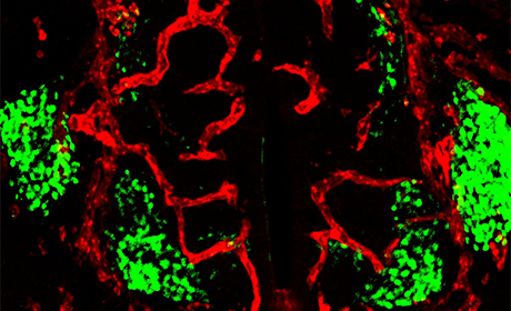
In their study, the Heidelberg researchers discovered that accurate vascularisation of the spinal cord during embryonic development is a highly precise mechanism, which requires a complexly regulated balance between growth-stimulating and growth-inhibiting factors. They demonstrated that spinal cord motor neurons produce the signaling molecule VEGF-A to stimulate the formation of blood vessels, and at the same time produce the VEGF-A trapping molecule sFlt1, which ensures accurate blood vessel patterning.
"Interestingly, even the slightest manipulation of this balance changes the pattern of blood vessel growth, causing the blood vessels to grow into the motor neuron region prematurely," explains the first author of the study, Patricia Himmels, a doctoral candidate in the research group led by Dr Ruiz de Almodóvar. The results of this study also suggest that premature vascularisation has a devastating impact on correct motoneuron positioning. These findings are particularly important for neurodevelopmental and neurodegenerative diseases, which also involve the vascular system. The Heidelberg researchers hope their results will help in the understanding of those disorders, and even perhaps lead to the development of new treatment strategies for these diseases.